Dumps AE-Adult-Echocardiography Collection & Guaranteed AE-Adult-Echocardiography Success
Wiki Article
DOWNLOAD the newest iPassleader AE-Adult-Echocardiography copyright from Cloud Storage for free: https://drive.google.com/open?id=1gWE_pDnBGubrDFOeoS33NHEMSXRnCeLp
iPassleader's ARDMS AE-Adult-Echocardiography practice exam software tracks your performance and provides results on the spot about your attempt. In this way, our ARDMS AE-Adult-Echocardiography simulation software encourages self-analysis and self-improvement. Questions in the ARDMS AE-Adult-Echocardiography Practice Test software bear a striking resemblance to those of the real test. This ARDMS AE-Adult-Echocardiography practice exam software is easily accessible on all Windows laptops and computers.
There are only key points in our AE-Adult-Echocardiography Training Materials. From the experience of our former customers, you can finish practicing all the contents in our training materials within 20 to 30 hours, which is enough for you to pass the AE-Adult-Echocardiography exam as well as get the related certification. That is to say, you can pass the AE Adult Echocardiography Examination exam as well as getting the related certification only with the minimum of time and efforts under the guidance of our training materials. So what you are waiting for? Just come and buy them!
>> Dumps AE-Adult-Echocardiography Collection <<
AE-Adult-Echocardiography - Pass-Sure Dumps AE Adult Echocardiography Examination Collection
Our AE-Adult-Echocardiography training materials provide three different versions to the client and they include the PDF version, PC version, APP online version. Each version’s using method and functions are different but the questions and answers of our AE-Adult-Echocardiography Study Materials is the same. The client can decide which version of our AE-Adult-Echocardiography exam questions to choose according their hobbies and their practical conditions.
ARDMS AE Adult Echocardiography Examination Sample Questions (Q45-Q50):
NEW QUESTION # 45
Which pathology is demonstrated in this video clip?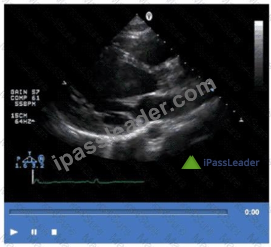
- A. Isolated left ventricular noncompaction
- B. Apical hypertrophic cardiomyopathy
- C. Amyloidosis
- D. Sarcoidosis
Answer: A
Explanation:
The video shows prominent trabeculations with deep intertrabecular recesses communicating with the left ventricular cavity, characteristic of isolated left ventricular noncompaction (LVNC). This congenital cardiomyopathy features a spongy myocardial appearance with thickened noncompacted layers.
Amyloidosis typically presents with thickened, bright myocardium but without prominent trabeculations.
Sarcoidosis involves granulomatous inflammation, and apical hypertrophic cardiomyopathy shows localized hypertrophy without trabecular changes.
This pathology is detailed in the "Textbook of Clinical Echocardiography, 6e", Chapter on Cardiomyopathies and Myocardial Disorders#20:360-365Textbook of Clinical Echocardiography#.
NEW QUESTION # 46
Which condition causes both tricuspid stenosis and tricuspid regurgitation?
- A. Carcinoid heart disease
- B. Amyloid heart disease
- C. Cor pulmonale
- D. Pulmonary hypertension
Answer: A
Explanation:
Comprehensive and Detailed Explanation From Exact Extract:
Carcinoid heart disease results from the deposition of fibrous plaques on the endocardium of right-sided heart valves, predominantly affecting the tricuspid and pulmonary valves. This leads to both tricuspid stenosis (valve leaflet thickening and immobility causing obstruction) and tricuspid regurgitation (incomplete coaptation due to leaflet retraction).
Pulmonary hypertension and cor pulmonale cause primarily functional tricuspid regurgitation without stenosis. Amyloid heart disease can cause restrictive cardiomyopathy but rarely causes combined tricuspid valve stenosis and regurgitation.
These pathological changes are detailed in the "Textbook of Clinical Echocardiography, 6e", Chapter on Carcinoid Heart Disease and Right Heart Valve Disease#20:335-340Textbook of Clinical Echocardiography#.
NEW QUESTION # 47
Which mitral valve filling pattern is characterized by a long deceleration time and an E/A ratio of 0.6?
- A. Pseudonormal
- B. Impaired relaxation
- C. Restrictive
- D. Normal
Answer: B
Explanation:
The mitral valve filling pattern characterized by a long deceleration time and a reduced E/A ratio (less than 1, such as 0.6) is consistent with impaired relaxation. This pattern is typically seen in early diastolic dysfunction, where there is slowed ventricular relaxation, resulting in reduced early diastolic filling (E wave) and a compensatory increase in atrial contraction contribution (A wave).
Impaired relaxation pattern shows:
E/A ratio < 1 (e.g., 0.6)
Prolonged deceleration time (>200 ms)
Prolonged isovolumic relaxation time (IVRT)
This pattern differs from restrictive filling, which has a high E/A ratio (>2), shortened deceleration time (<150 ms), and elevated left atrial pressures. Pseudonormal filling has a normal or near-normal E/A ratio but elevated filling pressures that mask underlying dysfunction and requires further evaluation with tissue Doppler or pulmonary venous flow for diagnosis. Normal filling has a typical E/A ratio around 1 to 1.5 with normal deceleration times.
The textbook details that impaired relaxation is the earliest sign of diastolic dysfunction and describes the prolongation of the deceleration time and reduced E/A ratio as hallmark findings of this stage.
NEW QUESTION # 48
Which of the following does the pulmonary capillary wedge pressure estimate?
- A. Left ventricular pressure
- B. Right atrial pressure
- C. Right ventricular pressure
- D. Left atrial pressure
Answer: D
Explanation:
Comprehensive and Detailed Explanation From Exact Extract:
Pulmonary capillary wedge pressure (PCWP) is obtained by advancing a balloon-tipped catheter into a small branch of the pulmonary artery and inflating the balloon to "wedge" the catheter, thereby occluding forward blood flow and measuring the pressure distal to the occlusion. The measured pressure reflects the pressure in the pulmonary venous system, which closely approximates left atrial pressure (LAP) under normal conditions.
Since the left atrium receives pulmonary venous return before the blood enters the left ventricle, PCWP is a surrogate for LAP, which in turn reflects left ventricular end-diastolic pressure (LVEDP) in the absence of mitral valve disease or pulmonary venous obstruction. PCWP is widely used in clinical and echocardiographic contexts to estimate left heart filling pressures.
It does not estimate right atrial, right ventricular, or left ventricular pressures directly. Right atrial pressure is measured via central venous pressure, right ventricular pressure by catheterization, and left ventricular pressure by direct catheterization.
This concept is extensively discussed in the "Textbook of Clinical Echocardiography, 6e", Chapter on Hemodynamics and Doppler Assessment, with specific emphasis on the use of PCWP to estimate left atrial pressure#20:200-210Textbook of Clinical Echocardiography#.
NEW QUESTION # 49
Which of the following can be calculated from the peak tricuspid regurgitant velocity?
- A. Mean pulmonary artery pressure
- B. Pulmonary artery diastolic pressure
- C. Right ventricular systolic pressure
- D. Right atrial pressure
Answer: C
Explanation:
Peak tricuspid regurgitant velocity (TRV) allows estimation of right ventricular systolic pressure (RVSP) using the simplified Bernoulli equation: RVSP = 4 × (TRV)
DOWNLOAD the newest iPassleader AE-Adult-Echocardiography copyright from Cloud Storage for free: https://drive.google.com/open?id=1gWE_pDnBGubrDFOeoS33NHEMSXRnCeLp
Report this wiki page